
Small Animal Imaging: University of Virginia Collaboration
A multimodality small animal imaging detector was developed to do cancer studies using live rats. Detectors were implemented that were able to image radiopharmacueticals that are used in SPECT (Tc99m) and PET (FDG).
 |
 |
| Digital x-ray detector. Cross sectional schematic of the six module FFDM detector showing two CCD modules (left side) and the readout electronics module (right side). | |
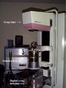
| Photograph of the dual-modality small animal imaging system at the University of Virginia. The compact hi-res gamma-ray detector was developed by Jefferson Lab. Brandeis University provided the digital x-ray camera. |
 |
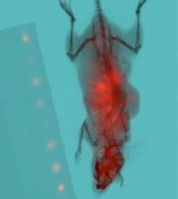 |
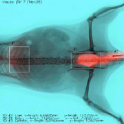
| Dual-modality mouse image from a dynamic Tc99m-pertechnetate study. The ROIs outlined indicate areas in which time-dependent radioactivity was measured. The ROIs are centerd on, the liver (left), colon (center) and catheter (right). | A co-registered FDG and x-ray image of a mouse with a human prostate tumor. |
 |
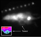 |
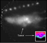
| Positron images of a mouse with human prostate tumor (left) and a mouse that has been transfected with VEGF (right). Note the absence of FDG uptake in the central portion of the tumor. | |