PEM Detector Development
Duke University Medical Center and Virginia Commonwealth University Health System
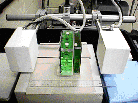
This detector prototype was designed for use in positron emission radiopharmaceutical studies of the breast. The two four inch square detectors allow a close imaging geometry and a small field-of-view to optimize the effectiveness of positron breast studies, see Figure 1. In order to evaluate the effectiveness of the system in cancer detection, we conducted a series of experiments to simulate patient imaging.
Simulation Experiments
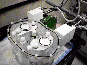
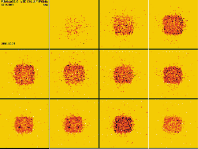
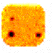
Using two plastic boxes to simulate the breast and small plastic spheres to simulate cancer lesions, we filled both with radioactive F18 labeled sugar water (Figure 2 Left). In addition, a large, multi-chambered vessel, called a human torso phantom, which mimics the position and shape of the internal organs was filled also filled with radioactive F18 labeled sugar water (Figure 2 Right). The concentration of radioactivity in the "torso", "breast" and "lesions" was typical of the concentrations one would find in patients injected with this radio-labeled sugar (F18 Fluorodeoxyglucose). We imaged this model with both a standard dual head Anger camera commercially available with positron detection software and with the Jefferson Lab dedicated positron imager. The resulting images are below.


