
Scintimammography
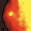
scan of a malignant breast tumor
Overview
The Detector and Imaging Group of the physics division at JLab has been collaborating with the University of Virginia in Charlottesville, VA, Hampton University in Hampton, VA and Dilon Technologies, Inc. in Newport News, VA on instrumentation projects to improve breast cancer detection.
Routine screening using x-ray mammography is currently the most sensitive method for the early detection of breast cancer. However, mammography can provide inadequate diagnostic information in women with radiodense breasts, and has limited ability to distinguish between benign and malignant lesions once they are detected. Imaging modalities with superior diagnostic specificity, such as nuclear imaging, using radionuclides producing single gamma rays (e.g. scintimammography) or emitting positrons (i.e. PET), are therefore needed to provide this additional information.
Scintimammography, like SPECT and PET nuclear medicine functional imaging, uses standard radiopharmaceuticals (radioactively labeled pharmaceuticals) to mark cancerous tissue with a gamma-ray emitting radioactive isotope. Typically a radiopharmaceutical such as Tc99m-Sestamibi is injected into a patient following a suspicious mammographic finding. The radiopharmaceutical propagates throughout the body via the bloodstream. The cancerous lesion has an increased metabolism (biological function) resulting in an increased accompanying uptake of the radiopharmaceutical. Hence the lesion can be detected some time after the injection by localization of "hot" spots of accumulated radioactivity within breast tissue. By placing gamma imaging detectors near the patient, an uptake map may be acquired that provides an indication of the likelihood that a suspicious mass is in fact a cancer.
Recently, interest in nuclear medicine detector development has focused on small, dedicated detectors for use in localization and diagnosis of breast cancer. Because of their small size, such new detectors can be placed closer to the areas to be imaged, and can be used in specialized applications or areas with limited space, where the use of a full-sized gamma camera is impractical or impossible. Such a system is referred to as a small field of view gamma imager.
The new compact JLab detector technology dedicated to breast cancer detection uses crystal scintillators to detect gamma-rays, as is the case with the standard SPECT and PET devices. Scintillators give off fast pulses of light (scintillation) when gamma-rays are detected in them. These light pulses are detected and converted into electrical signals by very sensitive light detectors called photomultiplier tubes. A major improvement in the new gamma detector technology used in the JLab application-specific breast imager (not found in standard nuclear medicine imagers) derives from the bright crystal scintillators being used in the form of a high resolution crystal array coupled to a high spatial resolution position sensitive photomultiplier (PSPMT). It is expected that this detector technology, when used in conjunction with standard mammography, will improve breast cancer detection, especially in cases where standard x-ray mammography is not sufficiently sensitive to provide early detection.
The major anticipated advantages of specialized small field of view scintimammography diagnostic devices may be summarized as follows:
- provide crucial functional and metabolic information not supplied by x-ray mammography
- improve efficiency, specificity and sensitivity relative to larger nuclear medicine devices and at a lower cost
- radiation doses delivered to patient can potentially be lowered
- combined with digital x-ray sensors for mammography, they can form the basis of a multi-modality system for breast imaging