
Scintimammography
A Combined Scintimammography/Stereotactic Biopsy Digital X-Ray System
Jefferson Lab, Hampton University and the Riverside Regional Medical Center are collaborating in a clinical study employing a dual modality imaging system utilizing scintimammography and digital x-ray. The purpose of the study is to obtain clinical data on the reliability of scintimammography in predicting the malignancy of suspected breast lesions with the ultimate goal to reduce the typically large (~65%) number of false positives associated with conventional x-ray mammography.
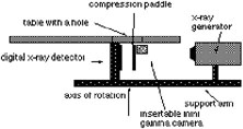
The scintimammography gamma camera is a custom built mini gamma camera with an active area of 5.3 cm x 5.3 cm based on a 2x2 array of Hamamatsu R7600-C8 position sensitive photomultiplier tubes (PSPMT). Optically coupled to the array of PSPMTs is a 16x16 matrix of NaI(Tl) crystal scintillators with each element 3 mm x 3 mm in size, separated by 0.3 mm and 6 mm thick. The compact gamma camera is attached to a Fischer Imaging Inc., digital x-ray stereotactic core biopsy system at Riverside Hospital's imaging clinic. The data acquisition system is based on a Macintosh G3 workstation which makes use of a four channel ADC PCI card. The final image overlay of the digital x-ray image and the gamma-ray image is achieved using the software application called IDL also running on the Macintosh computer.
 |
 |
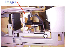
| small scintimammography mini-gamma camera attached to a stereotactic core biopsy system at Riverside Hospital. | |
 |
 |
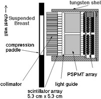
| Schematic diagram of the mini gamma camera detector head. | two of the PSPMTS and the NaI(Tl) crystal array with the lightguide in place. |
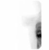
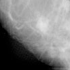
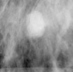
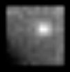
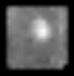
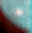
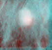
MiraLuma images of a positive breast lesion
| standard gamma camera | mini-gamma camera | digital x-ray | co-registered image |
|
|
|
|