
Small Animal Imaging: College of William and Mary Collaboration
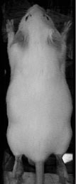
A high resolution detector system was used to image in a mouse the in vivo uptake of the iodine-125-labeled tracer methyl 3 b-(4-iodophenyl) tropane-2 b-carboxic acid methyl ester, also known as RTI-55(b-CIT). RTI-55 is used to study brain dopamine and seratonin transporters. The detector system is based on an array of CsI(Na) crystal scintillators coupled to a 125 mm diameter position sensitive photomultiplier tube. The project was supported in part by NSF grant # IBN9602865.
 |
 |
 |
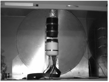
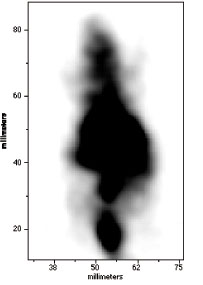
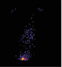
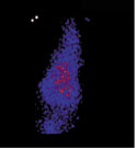
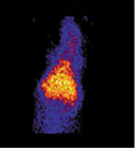
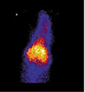
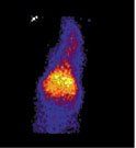
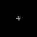
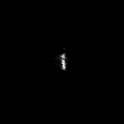
A photograph of the detector system with the rotating gantry mounted in a fume hood. The bottom detector is the Hamamatsu R3292 PSPMT with the CsI(Na) array and the top detector is the coincidence detector using a conventional 5 inch diameter photomultiplier tube. The middle image shows the biodistribution of RTI-55 in a 25 gm male CD-1 mouse. Total acquisition time of the image was 40 minutes which was started 30 minutes after a tail vein injection of ~15 mCi of 125I labeled RTI-55.
 |
 |
 |
 |
 |
 |
 |
 |
 |
|
 |
 |
 |
 |
|
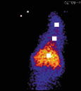 |
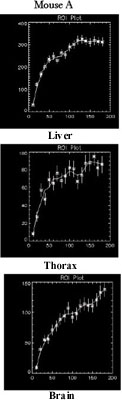
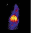
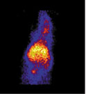
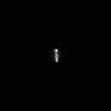
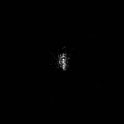
Images of bio-distribution of 2uCi of 125I-RTI-55 in an anesthetized mouse. Images and time activity curves are shown collected over the full body for a 360 minute period. The ROI plots from top to bottom are regions about 6 mm x 6 mm in the liver, thorax and brain. in which white squares indicate the locations of regions of interest (ROIs). | |||
 |
 |
 |
 |
 |
 |
 |
 |
 |
|
 |
 |
 |
 |
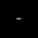
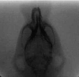
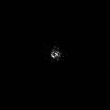
Digital x-ray imagetaken of a head of a mouse using a LIXI digital x-ray sytem. The sytem is being integrated into a gantry at the William and Mary Lab to do dual modality imaging with I-125. |
 |
 |
 |
 |
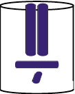 |
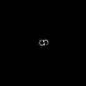
Twelve panels representing a sample of reconstructed tomographic slices from 60 projections of the phantom shown to the left. The blue regions indicate voids in a plastic rod. The phantom consists of four 30 mm long tubes, each of diameter 2 mm, drilled into a Lucite cylinder, 50 mm long by 32 mm in diameter. The voids were filled with approximately 20 uCi of radioactive iodine 125. |