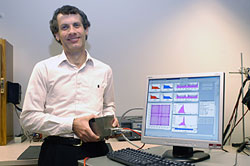
Mark Smith, Detector Group Biomedical Imaging Physicist and project manager for this effort, holds the tungsten box encasing the detector head for the mini gamma camera that Jefferson Lab's Detector Group built for the German Cancer Research Center. This photo was taken in December 2004 before the imaging device was disassembled for shipment to Germany. Final assembly is currently underway at the DKFZ.
Photo: Greg Adams, JLab.
This week three Jefferson Lab Detector Group members travel to Heidelberg, Germany, to assemble and bring on-line a small-animal imaging device the group developed and built for the German Cancer Research Center (DKFZ), an institute similar to the National Cancer Institute within the National Institutes of Health in the United States.
While nearly every member of the Detector Group played a role in developing or fabricating the device, Mark Smith, project manager for this effort, and Detector Group members Vladimir Popov and Ben Welch will be at the DKFZ during the week of April 24 to assemble, bring on-line and calibrate their mini gamma camera imager for small animals. Popov's primary responsibility is the camera's electronics, while Welch works on data acquisition and the user interface. He will also go over the user's manual with the scientists who will use the machine.
"In December we shipped the larger components and the electronics rack. During this trip we'll hand-carry the delicate electronics. Once there, our job will be to assemble the imaging device, get it up and running, and conduct a system calibration," Smith explained. "We'll also use this opportunity to teach the researchers how to use the camera and conduct their own calibration work."
Mastering the machine's capabilities and the calibration process will be critical to the cancer researchers' work as they will be using more than a dozen different radiopharmaceuticals in their research. Once assembled, the entire device will sit on a small, mobile cart and be integrated with an optical-imaging system to provide dual modality, small-animal imaging.
"We're very excited about getting the system operational," Smith said. "The imager will be used for research projects on a daily basis to gain physiological information on animal models of human disease."
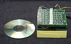
This close-up photo shows the key elements of the detector head (approximate size is 10 centimeters by 10 cm by 10 cm), which will fit inside of a tungsten box.
Photo: Greg Adams, JLab.
Jefferson Lab's work on the project began in June 2004. The Lab received approximately $86,000 from the DKFZ to build the gamma camera under a Work for Others Agreement. "This type of high-resolution device isn't available commercially and the research center didn't have the capability to fabricate its own," Smith explained. The basic device is along the same line as other small animal imaging gamma cameras developed by the Detector Group. However, the group is constantly refining their work, according to Smith. "This device has improvements; and we built it with a new type of flat panel, position sensitive photomultiplier tube," he explained.
The effort to develop international collaborations such as this one began a few years ago when Detector Group leader Stan Majewski tasked Mark Smith with finding potential scientific partners in Europe, to provide additional, crucial evaluations of Jefferson Lab designed imagers by implementing them in real biomedical projects. Smith traveled to Europe and after visiting several places in Netherlands and Germany, returned with the DKFZ collaboration plan. Smith's long-time friendship with a senior DKFZ scientist, Joerg Peter, was a plus in developing the working relationship. The two researchers have known each other since their years at Duke University Medical Center. "We stayed in touch over the years. Ideas grew up during our conversations during professional conferences, based on our mutual interests," Smith said. "He knew the sorts of things we could build here, so when they needed a small-animal imager, we were a good fit to build it for them."
The research institute plans to integrate the gamma camera with a non-JLab optical camera that will image bioluminescence and fluorescence markers for dual modality small-animal imaging, which will make it easier to correlate the information provided by the different imaging modes.
Jefferson Lab, or the Thomas Jefferson National Accelerator Facility, in Newport News, Va., is a basic physics research facility funded through the U.S. Department of Energy. Jefferson Lab's Detector Group assists with design and construction of apparatus for the highly complex spectrometers that gather subatomic particle data for nuclear physics experiments conducted at Jefferson Lab. The group has also used its core detector instrumentation expertise to develop a variety of medical and medical research imaging devices.
Relevant Images:
- Mini Gamma Camera Imager
- Mini Gamma Camera
- Detector Head
- Mark Smith, Detector Group Biomedical Imaging Physicist