Gamma Camera for a SPECT/Optical Small Animal Imaging System

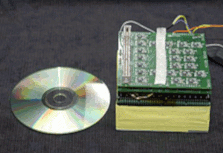
This close-up photo shows the key elements of the detector head (approximate size is 10 centimeters by 10 cm by 10 cm), which will fit inside of a tungsten box. Photo: Greg Adams, JLab.
Additional Links
Optical fluorescence and radiopharmaceutical imaging offer complementary ways of studying small animal physiology. A small, high resolution compact gamma camera has been developed and built at Jefferson Lab and integrated into a dual modality SPECT/optical small animal imaging system at the German Cancer Research Center (Heidelberg, Germany; Joerg Peter, PI). The key design features of the 10cm x 10cm field of view gamma camera are the use of a 2 x 2 array of flat panel position-sensitive photomultiplier tubes and a pixellated scintillation crystal array. A specially designed system of reflectors and mirrors built by the German Cancer Research Center permits simultaneous optical and gamma imaging with implicit image registration between the modalities. The optical detector is a high resolution cooled CCD camera. The optical and gamma subsystems are mounted on the same tomographic gantry. Initial small animal and phantom imaging studies have been performed to characterize the imaging system.
References:
Peter J et al., 2005 IEEE Nuclear Science Symposium Conference Record. Bo Yu, Ed. Fajardo, Puerto Rico, October 23-29, 2005. IEEE Catalog Number 05CH37692C, pp. 1969-1972

