Triple Modality Small Animal Planar Imaging System

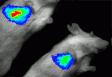
This false-color image of stem cells inside mice was captured by the cooled CCD camera/optical modality of the small animal planar imaging system developed for Case Western.
Additional Links
Jefferson Lab in collaboration with Case Western Reserve University (Dr. Zhenghong Lee) is developing a system that provides fused planar radiopharmaceutical imaging, planar x-ray imaging and planar bioluminescent/fluorescence imaging in a single apparatus for small animal imaging. Jefferson Lab developed a high resolution gamma camera based on a large position sensitive photomultiplier tube coupled to a pixellated NaI(Tl) scintillator array with individual crystal elements 1.3 mm x 1.3 mm x 6 mm in size and 0.2 mm septa between each element (1.5mm pixel step). The gamma camera has an active area of ~10 cm in diameter and has been optimized for I-125 imaging. To acquire anatomical information we are using a commercial digital x-ray detector that provides a field of view of 5 cm x 10 cm. A cooled CCD based camera is utilized for the imaging of the bio-distribution of bioluminescence. All three imaging instruments are to be integrated into a single light tight / x-ray tight enclosure. The system is being used for several mouse studies such as Afp based liver cancer imaging, cystic fibrosis gene therapy, new peptide ligands targeting αvβ3 for angiogenesis and new small-molecule-based radiotracers for prostate and other cancers.
References:
Weisenberger AG et al., 2005 IEEE Nuclear Science Symposium Conference Record. Bo Yu, Ed. Fajardo, Puerto Rico, October 23-29, 2005. IEEE Catalog Number 05CH37692C, pp. 1761-1764
Martone PF et al., J Nucl Med. 2004; 45: 421P [abstract]

