|
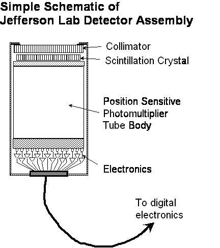
Once through the collimator, photons strike a sodium iodide scintillation crystal. The crystal absorbs the gamma radiation photons and converts them into light photons that can be detected by a photomultiplier tube. Exiting the crystal, the light photons enter a position sensitive photomultiplier tube, where the light signals are detected, amplified and converted into electrical pulses. The pulses are then digitized and sent to a computer where sophisticated software translates the digital signal into an image. Click image for larger file (9KB)
|
Newport News, VA. — To study the structure of the nucleus of the atom, DOE’s Thomas Jefferson National Accelerator Facility develops and employs a wide range of cutting-edge detector technologies. Now, Jefferson Lab scientists have used their expertise to build a small animal medical imager that’s helping researchers develop a new gene therapy technique for cystic fibrosis.
“Our core expertise is instruments. Once you have instruments with different imaging capabilities, you want to spin-off that technology in ways that can do the most good,” says Stan Majewski, Jefferson Lab Detector Group leader.
For use in new, more sensitive detectors for medical imaging, Majewski and his group are adapting the technology that detects photons and other high-energy particles produced when the Lab’s electron beam strikes an experimental target. In medical imaging, photons are emitted by a radioactively labeled molecule that has attached to other molecules in the body. This technology allows doctors to image cellular function in areas of interest and has already proved its usefulness, for instance, in imaging cancer tumors.
According to Jefferson Lab’s principal investigator on this project, Drew Weisenberger, the team has designed and built several small animal imagers for use in biomedical research. “Researchers use animals all the time in developing imaging technologies. Dr. Lee is using our technology in research with mice, and we’re working together with him to improve the technology and produce the images he needs,” Weisenberger says.
Zhenghong Lee, a researcher with Case Western Reserve University School of Medicine and University Hospitals of Cleveland, is using the small animal medical imager in cystic fibrosis gene therapy studies along with colleagues Assem Ziady and Pamela Davis. They’re researching a new way to replace the defective gene that causes cystic fibrosis, a deadly disease that affects about 30,000 Americans and which has no known cure. In conventional gene therapy, a virus is used to deliver a therapeutic gene to the target cells.
|
|
But this method isn’t viable for cystic fibrosis patients, who are already battling chronic lung infections and inflammation. “If we deliver a virus to the lungs, even if it’s a hollow virus, it will initiate an inflammatory response, making the patient even sicker. So that’s the last thing we need in the cystic fibrosis patient,” Lee says.
Lee and his colleagues hope to solve this problem by delivering gene therapy through an aerosol that patients can inhale. In a previous study, he found that dripping a solution containing a functioning copy of the cystic fibrosis gene (CFTR) into the noses of mice without the gene could successfully deliver it into the lungs.
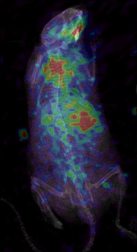
Successful gene therapy depends not only on where genes are delivered, but also on how well genes are transferred — the ability of cells in the body to take and express the new gene as one of their own. Recently, Lee and his colleagues repeated the experiment with HSV1-tk, a gene often used to measure the expression of transferred genes. A day later, the experimenters injected a radioactive tracer and acquired x-ray and gamma images of the mice with Jefferson Lab’s custom-built small animal imager — a planar gamma scintigraphy device. The study revealed that the mice cells were expressing the new gene in the lungs and that Jefferson Lab’s small animal imaging system can successfully image gene transfer with nuclear imaging techniques.
Lee says the next step is to continue to improve Jefferson Lab’s small animal imager to get even clearer measures of gene function after inhaled gene therapy. “We want to introduce a third imaging modality, bioluminescence, which can be measured with an optical camera. These imaging methods will be independent and cross-check each other,” Lee says.
Weisenberger agrees. “I think a tri-modal imaging device has the potential to bring a very powerful tool to biomedical research. The first modality, x-ray, tells you about structure. The other two modalities, gamma emission and optical, can both give you information on function, allowing doctors to measure two different functions at the same time,” he explains.
Lee says that he wants to develop and test the inhalation gene therapy technique to prove its safety and effectiveness in small animals, and eventually, he wants to use a therapeutic CFTR gene with the therapy technique in patients. “The research in small animals is just preliminary work. Ultimately, we want to go to clinical trials in humans. Right now, cystic fibrosis is a fatal disease. But if everything works out, and we can make it a manageable disease, I think that would be really, really good,” Lee says.
|
|

The CFTR gene codes for a protein that controls the transport of chloride ions into and out of individual cells in the lungs, liver, pancreas, digestive tract, reproductive tract and skin. The most common CFTR defect causes the body to make a defective protein that is destroyed by the body. As a result, chloride and sodium levels become imbalanced, leading to abnormally thick mucus. This mucus clogs the lungs, trapping bacteria and causing recurring lung infections; it also prevents digestive enzymes from reaching the intestines to break down and absorb nutrients from food. Current treatments are prolonging lives but haven’t conquered the disease; most patients eventually succumb to lung disease.
For more information, or to schedule an interview, contact:
Linda Ware
ware@jlab.org
phone (757) 269-7689
fax (757) 269-7398
Kandice Carter
kcarter@jlab.org
phone (757) 269-7263
fax (757) 269-7398
Front page image: Zhenghong Lee, Case Western Reserve University School of Medicine and University Hospitals of Cleveland, and Drew Weisenberger, Jefferson Lab, with an image showing successful gene transfer.