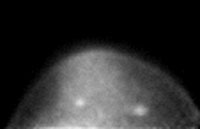
This PEM image shows two cancerous lesions. The one on the right was depicted by conventional mammography, but the one on the left was only identified by the PEM unit.
Image courtesy: Eric Rosen, Duke University Medical Center
Newport News, VA — A study published in the February issue of the journal Radiology shows that a positron emission mammography (PEM) device designed and built by Jefferson Lab scientists is capable of imaging breast cancer tumors. In the pilot study, conducted by Duke University Medical Center researchers, the unit imaged 18 malignant tumors in 23 patients receiving additional screening due to suspicious mammograms.
For many women, regular mammograms allow physicians to spot breast cancer tumors as dense lumps in the breast. But mammography often fails in women who have dense breast tissue due, for instance, to genetics or scarring. According to Eric Rosen, M.D., a Duke University Medical Center physician and lead author on the study, "In women with dense breasts, it's very hard to pick out even large anatomic abnormalities."
Stan Majewski, Jefferson Lab Detector Group Leader and principal investigator on the instrumentation part of the project, led the team that designed and built the PEM unit. He says PEM imaging works differently than mammography. It reveals breast tissue that is showing higher metabolism than other areas. "The imager we built is a functional imager. That is, it indicates something about physiology, which can be different from anatomy," he says.
To fuel rapid growth, cancer cells use more glucose (sugar) than surrounding cells. In this imaging procedure, a small dose of radioactive molecules that look like sugar, called fluorodeoxyglucose (FDG), are injected into the body, where they're absorbed by cancerous tumors. The PEM device pinpoints tumors in the breast by detecting the location of FDG uptake. "By detecting areas that have increased glucose metabolism, you can often distinguish a cancer between normal surrounding tissue, which in general has low uptake of FDG," Dr. Rosen says.
For the study, Duke physicians recruited patients with suspicious mammograms who were scheduled for biopsies. "We recruited 23 patients that had 23 lesions that were highly suggestive of malignancy. PEM showed 20 lesions, 20 abnormalities, of which 18 were cancer and 2 were not cancer," Dr. Rosen says. The PEM unit missed three tumors, all of which were located very close to the chest wall, an area that PEM doesn't image well. And of the 20 lesions spotted by the PEM system, one was not picked up by mammography. A subsequent biopsy revealed that this additional lesion was cancerous.
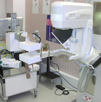
The PEM unit (on the left) was designed and built by Jefferson Lab scientists. A recent study proved the unit is capable of imaging breast cancer tumors. A standard x-ray mammography unit sits to the right of the PEM device.
Image courtesy: Tim Turkington, Duke University Medical Center
"We wanted to make a difference with this imager. Our expertise in building detectors for Jefferson Lab's nuclear physics program allowed us to build a device that's sensitive to the presence of the radioactive molecule, FDG," Majewski says, "And now we're seeing the results of that. They detected an additional lesion that was not on mammography. That can directly impact patient care."
Dr. Rosen says the study was indeed a success, "What we concluded is that our PEM unit is capable of detecting cancer, it's capable of demonstrating small breast malignancies, and that it can be performed in the breast clinic with a small dose of FDG and a very short, 5-minute acquisition time."
The Jefferson Lab/Duke team is now modifying the PEM system and imaging procedure to allow for better detection of lesions located near the chest wall. Dr. Rosen says the next step is to figure out what size and types of cancer tumors the unit is best capable of detecting, exactly how sensitive the unit is, and where it fits in the cancer screening process. "So now what we want to do is study a larger population and study a more representative population of patients," he says. The National Cancer Institute has funded a larger study that will include 200 patients to begin answering those questions.
More information: