NEWPORT NEWS, Va. -- The positron emission mammography/tomography breast imaging and biopsy system was designed and constructed by scientists at Jefferson Lab, West Virginia University and the University of Maryland School of Medicine. The PEM/PET system is designed for detecting and guiding the biopsies of suspicious breast cancer lesions.
"This is the most-important and most-difficult imager we've developed so far," Stan Majewski, Jefferson Lab Radiation Detector and Medical Imaging Group leader says.
How It Works
The PEM/PET system features components designed for imaging the unique contours of the breast. The complete system is the brainchild of Ray Raylman, a professor of radiology and vice chair of Radiology Research at WVU. He says the technology is built on traditional PET imaging, where a drug with a radiation-emitting component is injected into a patient and is then imaged. The system is designed for imaging tumors in women who have indeterminate mammograms because of dense or fibroglandular breasts.
"I got the idea as a postdoc when I was working with some radiologists who wanted to use PET to image breast cancer. They could do it, but they couldn't get the resolution," Raylman says.
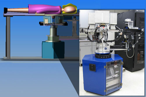
Schematic drawing of the PEM/PET imaging and biopsy system. Image: Ray Raylman
In this imaging procedure, a small dose of radioactive molecules that look like sugar, called fluorodeoxyglucose (FDG), are injected into the body, where they're absorbed by cancerous tumors.
"The patient lays on the bed, with one breast at a time falling through a hole in the table that goes to the center of the scanner, which is sort of the standard way to do stereotactic breast biopsies right now," Raylman explains.
The system images the breast with a movable array of two pairs of two flat scanners. The scanners detect the radiation from the drug, and a sophisticated software package produces a three-dimensional image from scanner information. The images are available in about 10 minutes from the start of the scan.
If a suspected lesion is found, a single pair of scanners is then used to guide a needle biopsy of the lesion. The biopsy is performed with a person-controlled robot arm.
"Just before we take the tissue sample, we take a new image. The needle we use has a little radioactive source embedded in its tip, so that we can see the relative position between the biopsy needle and our suspicious lesion," Raylman says.
A Difficult Task
One important difficulty to overcome was the time it takes for the system to combine the information of the individual scanners into a three-dimensional image.
Mark Smith is currently an associate professor in the Department of Radiology at the University of Maryland School of Medicine. While working on the software, he was affiliated with both the Jefferson Lab Radiation Detector and Medical Imaging group and UM.
"I developed the image reconstruction algorithms so that we can use the data from the detector system to form a three-dimensional image. And this is challenging, due to the fact that we want image reconstruction to be done within approximately five to ten minutes after the woman has been imaged with the device," he says.
System Testing
Raylman led the initial testing of the system and is the lead author on the study. Raylman's team imaged various radioactive sources to test the resolution of the system. In one test, a series of capillary tubes were imaged to test the resolution capabilities.
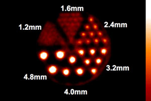
PEM/PET image of rods inside a plastic breast phantom show the resolution capabilities of the device. The diameters of the rods (in millimeters) are shown. Technical aspects of the image may be found in the paper, cited below. Image: Ray Raylman
To visually demonstrate the capabilities of the scanner, fluorine-18, a radioactive source, was deposited in thin rods of known diameter inside a plastic device used as a proxy for the breast – a breast phantom. In another test, two spheres of radioactive material in a water-filled cylinder, another type of breast phantom, were also imaged.
"We had good performance characteristics, with image resolution below two millimeters. In regular PET, the image resolution is over five millimeters, so we're quite a bit better than that," Raylman says.
In addition, thanks in part to speedy image reconstruction, the initial tests revealed that the PEM/PET system takes about the same amount of time to image cancer and complete a biospy in about the same amount of time as a traditional biopsy.
The Next Steps
The next steps for the team include minor improvements in the detector systems and image reconstruction software and the addition of components for taking x-ray computed tomography (CT) scans. Initial clinical trials are planned after completion of system testing.
"The system is another example of nuclear physics detector technology that we have put a lot of time and effort into adapting for the common good," Majewski says.
Smith agrees. "I'm continuing to collaborate with Jefferson Lab and Ray Raylman on this particular project, because I feel very strongly it's something I'd like to see clinically implemented."
Other Radiation Detector and Medical Imaging group members that worked on the project include Brian Kross, John McKisson, Vladimir Popov (now with the Jlab Radiation Control group), James Proffitt and Drew Weisenberger.
The work was supported by a National Cancer Institute grant to West Virginia University and by DOE's Office of Science. Construction of the Jefferson Lab portion of the imager was supported through a subcontract from WVU.
The results of these tests will be published in the journal Physics in Medicine and Biology on Feb. 7.
Technical Paper:
The positron emission mammography/tomography breast imaging and biopsy system (PEM/PET): design, construction and phantom-based measurements
Raymond R Raylman, Stan Majewski, Mark F Smith, James Proffitt, William Hammond, Amarnath Srinivasan, John McKisson, Vladimir Popov, Andrew Weisenberger, Clifford O Judy, Brian Kross, Srikanth Ramasubramanian, Larry E Banta, Paul E Kinahan and Kyle Champley
Further Reading:
Nuclear Physics Technology Saves Lives
Jefferson Lab Medical Imager Spots Breast Cancer