NEWPORT NEWS, Va. -- When Sue Parham underwent tests prior to undergoing surgery for breast cancer, she had no idea how much her life was about to change.
Parham's surgeon had sent her to the hospital for a new procedure designed to provide better images of a small tumor in her left breast. To get those better images, Parham underwent a BSGI scan, short for Breast-Specific Gamma Imaging, at Legacy Good Samaritan Hospital in Portland, Ore.
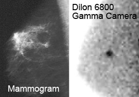
The Dilon 6800 Gamma Camera uses technology originally developed for basic nuclear physics research to image breast cancer in women. These sample images are of a patient with a history of breast cancer in the left breast. The mammogram of the patient's right breast shows an area of dense tissue that was considered normal but was being monitored in yearly mammograms. The Dilon gamma camera image reveals the cancer missed by the mammogram, probably saving this patient's life.
Based on gamma imaging technology developed at the U.S. Department of Energy's Jefferson Lab, BSGI is a non-invasive breast imaging procedure that captures the cellular function of breast tissue, complementing mammography in helping to resolve difficult to interpret cases.
"As the fifth patient to use the Breast-Specific Gamma Imaging at Portland's Good Samaritan Hospital, I could tell they were still working with the technology. But that didn't explain what was to follow," the 49-year-old Parham recalled of the procedure she underwent a year ago.
The BSGI found "a slow-growing, potentially deadly tumor in my right breast that had reached 3 centimeters in size that had gone unnoticed by the mammogram," Parham said. And it was then discovered that the cancer had spread to the lymph nodes in her right arm.
"My diagnosis moved from straightforward to complex, my treatment from routine to aggressive and my life from normal to anything but," Parham explained.
"For me, the past year has been the most challenging of my life. I've endured a double-mastectomy, removal of the lymph nodes under my right arm, removal of both ovaries, reconstructive breast surgery, four-and-a-half months of chemotherapy, and six-and-a-half weeks of radiation therapy. But, most important, I am alive. I am a living, breathing testament to the early detection made possible by BSGI."
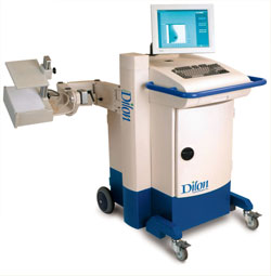
The BSGI machine used to diagnose Parham's cancer was manufactured by Dilon Technologies, a Newport News company. Dilon licensed its high-resolution gamma imaging technology from Jefferson Lab after recognizing its potential for use in marketable compact dedicated breast imager camera.
The patented imaging technology was developed by Jefferson Lab's Radiation Detector and Medical Imaging Group. The group began exploring the use of nuclear physics detector technology for medical imaging applications in 1995, starting with two types of breast imagers. Members of the group are experts in radiation detector technology, which is used extensively at Jefferson Lab to conduct basic nuclear physics research. In the biomedical field, the group has been involved in the development of high-resolution gamma and positron imagers and in novel multimodality (including X-ray and optical) imaging concepts.
"It's a never-ending challenge and desire for us to take our technical expertise and make a difference in people's lives. BSGI is the first of what we anticipate will be still more instruments we hope to develop and help bring to market," says Stan Majewski, head of Jefferson Lab's Radiation Detector and Medical Imaging Group.
BSGI is designed for situations where mammography is inconclusive and further evaluation is needed, especially when patients have dense breast tissue, implants, multiple suspicious lesions or clusters of microcalcifications, palpable lesions not detected by mammography or ultrasound, post-surgical or post-therapeutic mass, or if they have been taking Hormone Replacement Therapy.
Parham had dense breast tissue, and she noted, "I didn't know there was a connection between breast density and breast cancer, nor did I understand that mammograms were less effective on women with dense breasts."
The link is that both cancer and breast tissue are dense in nature, and the more dense the breast tissue, the more likely that a cancer could be missed in evaluation, especially with imaging tests that look at structure and not. metabolic function, such as mammography and ultrasound.
In the Newport News area, BSGI proceedures are available at Riverside Diagnostic and Breast Imaging Center and the Dorothy G. Hoefer Comprehensive Breast Center at Sentara Port Warwick. Dr. Curtis Stoldt, director of Riverside Diagnostic and Breast Imaging, says he first heard about BSGI in 2005 at a conference and immediately began working to secure a BSGI machine for his hospital.
"It took three years but it is online and we are doing 6-8 patients a week, and that number is increasing quickly," Stoldt says.
"BSGI images are simpler and easier for the patient and technologist. The studies are much simpler to read and compare with mammograms," Stoldt adds. "There are fewer studies with questionable or indeterminate findings, and my overall confidence is greater."