Large Field of View Positron Emission Mammography Imaging Devices

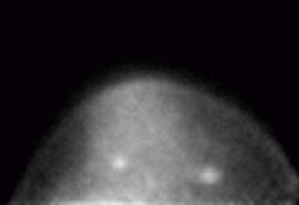
This PEM image shows two cancerous lesions. The one on the right was depicted by conventional mammography, but the one on the left was only identified by the PEM unit. Image courtesy: Eric Rosen, Duke University Medical Center
Additional Links
Dedicated positron emission mammography (PEM) systems potentially provide a high sensitivity, high resolution alternative to whole body PET for positron breast imaging. In collaboration with Duke University Medical Center (Tim Turkington, PI), we have designed, built and evaluated a large field of view (15 cm x 20 cm) PEM system. The device is built with a set of two pixellated LGSO/LYSO crystal scintillators coupled to arrays of compact position sensitive photomultiplier tubes. In pre-clinical trials performed at Duke lesions as small as 4 mm were seen in phantom experiments using advanced iterative image reconstruction algorithms developed at Jefferson Lab. Subsequent clinical trials at Duke supported by the DoD (Eric Rosen, PI) showed that small primary breast malignancies can be imaged. An NIH-funded clinical study at Duke is underway to scan 200 patients with suspected breast cancer (Mary Scott Soo, PI).
References:
Rosen EL et al., Radiology. 2005; 234: 527-534
Turkington TG et al., 2002 IEEE Nuclear Science Symposium Conference Record. Scott Metzler, Ed. Norfolk, Virginia, November 10-16, 2002. ISBN 0-7803-7637-4

