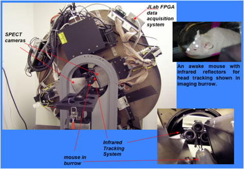
The Radiation Detector and Imaging Group with involvement from numerous collaborators has developed pre-clinical radiation imaging systems for small animal research based on positron emission tomography (PET) and single-photon emission computed tomography (SPECT). The group in collaboration with Oak Ridge National Laboratory, Johns Hopkins University and the University of Maryland are developed a SPECT-CT system for imaging unanesthetized, unrestrained mice. The use of anesthesia and physical restraint can change the neurological and physiological processes being studied. With the system the position and pose of the head of the unanesthetized mouse is recorded during list mode SPECT image acquisition. The gamma-ray image data is later registered to the time-varying animal orientation.
Motion Tracking Enabled Radioisotope Imaging/Radioisotope Imaging Detectors
The Jefferson Lab Radiation Detector and Imaging Group with Oak Ridge National Laboratory and Johns Hopkins University built a medical imaging system using nuclear physics detector technology to allow medical imaging without the use of anesthesia. The technology is a novel implementation of single photo emission computed tomography (SPECT). This provides a unique imaging tool for basic research into human diseases such as Alzheimer's and Parkinson's disease. The technology also has applications in pediatric imaging and in cases in which a patient cannot remain motionless. Recently, the collaboration used the system to investigate the effect of anesthesia on the uptake of 123I-DaTSCAN™ in mouse striatum. DaTSCAN™ is a dopamine transporter (DaT) ligand containing ioflupane (123I) INN, which is a radioiodinated cocaine analog with high affinity to DaT located on the pre-synaptic nerve endings in the striatum. It is also indicated to help differentiate probable dementia with Lewy bodies (DLB) from Alzheimer's disease.
|
Awake Animal SPECT system at Johns Hopkins University The SPECT data were reconstructed using 2D-OSEM. SPECT and x-ray CT images were co-registered manually. Regions of interest were drawn in left and right striata (STR), and cerebellum (CER), and the tracer binding potential (BP) was calculated as STR/CER-1. Outcomes of the 2nd scan (45-75 minutes post-injection), showed there was over a 30% statistically significant decrease seen in the awake mice. This study demonstrated significantly higher uptake of DaTSCAN™ in striata of anesthetized mice as compared to awake mice, indicating that anesthesia can have a substantial effect on tracer studies. |
 |
|
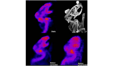 |
Awake Animal SPECT system at Johns Hopkins University |
|